2018, University of Montevallo, Killian's review: "Rythmol 150 mg. Trusted online Rythmol.".
The dermal-epidermal junction is highly convoluted ensuring a maximal contact area purchase rythmol 150 mg otc. Other anatomical features of the skin of interest are the appendageal structures: the hair follicles buy rythmol 150mg cheap, nails and sweat glands generic 150mg rythmol with amex. The keratinocytes comprise the major cellular component (>90%) and are responsible for the evolution of barrier function. The epidermis per se can be divided into five distinct strata which correspond to the consecutive steps of keratinocyte differentiation. The ultimate result of this differentiation process is formation of the functional barrier layer, the stratum corneum (~0. The stratum basale or basal layer is responsible for the continual renewal of the epidermis (a process occurring every 20–30 days). Proliferation of the stem cells in the stratum basale creates new keratinocytes which then push existing cells towards the surface. The next layer of the epidermis is the stratum spinosum, named for the numerous spiny projections (desmosomes) on the cell surface. The keratinocytes maintain a complete set of organelles and also include membrane-coating granules (or lamellar bodies) which originate in the Golgi. Subsequently, we encounter the stratum granulosum or granular layer, characterized by numerous keratohyalin granules present in the cytoplasm of the more flattened, yet still viable, keratinocytes. More lamellar bodies are also apparent and concentrate in the upper part of the granular cells. The transition layer, the stratum lucidum, comprises flattened cells which are not easy to visualize microscopically. The cellular organelles are broken down leaving only keratin filaments in the stratum granulosum an interfilament matrix material in the intracellular compartment. The membrane coating granules fuse with the cell membrane and release their contents into the intercellular space. Finally, in the stratum corneum, the outermost layer, protein is added to the inner surface of the cell membrane to form a cornified envelope that further strengthens the resistance of the cell. A layer of lipid covalently bound to the cornified envelope of the corneocyte contributes to this exquisite organization. The intercellular lipids of the stratum corneum include no phospholipids, comprising an approximately equimolar mixture of ceramides, cholesterol and free fatty acids. These non-polar and somewhat rigid components of the stratum corneum’s “cement” play a critical role in barrier function. On average, there are about 20 cell layers in the stratum corneum, each of which is about 0. Yet, the architecture of the membrane is such that this very thin structure limits, under normal conditions, the passive loss of water across the entire skin surface to only about 250 mL per day, a volume easily replaced in order to maintain homeostasis. For example, changes in intercellular lipid composition and/or organization typically result in a defective and more permeable barrier. Skin permeability at different body sites has been correlated with local variations in lipid content. And, most convincingly, the conformational order of the intercellular lipids of the stratum corneum is correlated directly with the membrane’s permeability to water. Taken together, it has been deduced that the stratum corneum achieves its excellent barrier capability by constraining the passive diffusion of molecules to the intercellular path. This mechanism is tortuous and apparently demands a diffusion path length at least an order of magnitude greater than that of the thickness of the stratum corneum. Thus, the stratum corneum is most convincingly viewed as a predominantly lipophilic barrier (this makes perfectly good sense as it was designed to inhibit passive loss of tissue water in an arid environment), which manifests a high degree of organization, and which constrains permeating molecules to a long and convoluted pathway of absorption. These characteristics dictate the permeability of the membrane and determine the extent to which drugs of various physicochemical properties may be expected to transport. The extensive microvasculature network found in the dermis represents the site of resorption for drugs absorbed across the epidermis; that is, it is at this point that transdermally absorbed molecules gain entry to the systemic circulation and access to their central targets. The dermis also supports skin’s appendageal structures, specifically the hair follicles and sweat glands. With respect to drug delivery, interest in these structures has centered upon the possibility that they may provide “shunt” pathways across the skin, circumventing the need to cross the full stratum corneum. However, surface area considerations mean that the appendages cannot contribute significantly to the overall drug flux. Transdermal bioavailability therefore and strategies to improve delivery often involve changing the composition or the organization of the intercellular lipids. Such enhancing technologies are of course feasible, but not without problems (see below). For very lipophilic compounds (say, those with octanol-water partition coefficients greater than 10 ), it is generally believed4 that transport is limited not by diffusion across the stratum corneum, but rather by the kinetics with which the molecule leaves this membrane and enters the underlying (and much more aqueous in nature) viable 193 epidermis. Compounds exhibiting this behavior also manifest two other problems with respect to transdermal bioavailability. First, the “lag-time” observed prior to their appearance at useful levels in the blood may be significantly prolonged by the slow partitioning kinetics (see Figure 8. Second, these substances, because of their strong attraction for the lipophilic environment of the stratum corneum, often form significant reservoirs in the membrane from which release may continue even after removal of the delivery system. In fact, there is variability but, over most of the surface, this is not greater than the normal inter-individual variability observed at a specific site. Certain regions are significantly more permeable—the genitalia, especially the scrotum, the axilla, the face, the scalp, and post-auricularly.


In some cases the indication for a special remedy purchase rythmol 150 mg amex, like one of these buy 150mg rythmol amex, is so marked purchase rythmol 150 mg visa, that we give it alone, and it quickly cures most severe and obstinate diseases. I would like to continue this subject further, for it is one in which I am greatly interested, and I know it is one in which you are interested, but the shortness of our session will not permit further remarks. But when we come together another year, with another year’s experience, we may discuss it again. The practice of medicine is proverbially uncertain, not so much possibly as regards the termination of disease, as regards the influence of medicine to palliate or arrest it. Instead of making this uncertainty a cardinal doctrine, a belief in which is absolutely essential to regularity, it seems to me it would be profitable to examine it carefully, and by analysis determine the “elements of uncertainty;” we might then hope to determine the “elements of certainty,” and by a simple process of reasoning, avoid error and attain truth. The most important factor in “medical uncertainty,” is undoubtedly our present nosology. The element of uncertainty lies here, that a name employed to designate a disease, may cover the most diverse pathological states. The case of to-day, and the case of to-morrow, though justly called by the same name, may require a widely different treatment; the remedies employed successfully in one, would increase the disease in the other. Every one of our readers may draw the evidence of the truth of these propositions from his own practice. The second “element of uncertainty” we find in the doctrine of idiosyncrasy, which is also a cardinal article of faith. We are gravely taught that in medicine one of nature’s laws - that “like causes produce like effects,” is inoperative; and, on the contrary, “that no man can possibly tell from the action of a medicine on one, what will be its influence upon another. The third “element of uncertainty” lies in the application of the Latin motto, post hoc ergo propter hoc - that which follows a medicine must be due to its influence. If a man recovers from sickness after taking Podophyllin or Quinine, it is due to these agents, and he probably would not have gotten well without them. But there is this singular fact here: whilst physicians are willing to credit their remedies with all relief from suffering, improvement and restoration to health, they are not willing to reverse it, and concede increase of suffering, prolongation of disease and death to the remedies, though the sequence is quite as natural in the one case as the other. It does seem strange that physicians should have so thoroughly believed that medicine saved the lives of the sick, that without it the majority, or all, would have died. Even now when all this is proven beyond cavil, by some of the best observers, we find the majority won’t believe it; even if they concede it in theory, they deny it in practice. The fourth element of uncertainty lies in the endeavor to get direct results from indirect agencies. You want to influence the circulation and the temperature, and you give remedies which produce emesis or catharsis. You want to elect Greeley, and you whip your neighbor because he “rah’s” for Grant. The fifth element of uncertainty is the administration of remedies in poisonous instead of medicinal doses. It is true that the poisonous action may be known with some certainty, but its influence upon disease is very uncertain. The poisonous action sets up a new process of disease in so far as it changes structure and function, and it may be curative according to the law of substitution. A medicinal action we understand to be one that restores function, and thus removes disease. Give your remedy in medicinal doses, and then you may expect direct and positive results in the relief of disease. These are the principal elements of uncertainty in regular medicine and in our school. To these we might add a number of minor ones, among which is a belief in “special providences,” inscrutable or otherwise. Indeed any one who is inclined to shift his responsibility upon his Creator, and to believe that the laws of nature can be suspended from any providence, has all the elements of uncertainty with him. The principal element of uncertainty in Homœopathic medicine is the making of pain a principal symptom, and the treatment of symptoms in place of pathological conditions. The cheerful trust in Nature of the high dilutionist is laudable, though we can’t say so much for their claim to all the glory of relief and recovery. This is but a rough sketch of the subject, which we present as good material for thought. In our next article, we will consider the “elements of certainty” in medicine, in the same order. In our last article we briefly discussed the “elements of uncertainty in medicine,” and we now propose to look at the other side - how may we attain certainty in medicine. We all agree that the practice of medicine in the past has been notoriously uncertain, and that there is yet great room for improvement. The first, and most important element of “uncertainty” is found in our present nosology, and the constant tendency to prescribe for names of disease. The first “element of certainty” will be, therefore, an entire avoidance of this error, diagnosing pathological conditions and prescribing for these. We have heretofore seen that disease, as we meet it in the individual, consists of a series of functional lesions - all disease is an impairment of function. Certain prominent lesions or symptoms give a name to the disease, according to the present nosology, but the name does not, and can not convey to the mind the character of the lesions or the treatment required to remove them. If I say my patient has “pneumonia” (taking one of the simplest diseases), I give you no information which would guide you to a correct treatment, and if you prescribe it must be upon the idea that all inflammations of the lung are alike, unless you follow the expectant plan, and use the “mush poultice,” with rest and good nursing. In one case the prominent lesion would be of the circulation and temperature; and we would stop the inflammation by the third day with the use of Veratrum and the bath, alone. In a third class of cases, with especial impairment and feebleness of mucous structure, Ipecac would be a prominent remedy. In a fourth class of cases, with special impairment of skin and dryness of mucous membranes, we would use Asclepias. In a fifth, if we had the broad pallid tongue, we might treat the case with Bicarbonate of Soda alone.

Chronic renal failure Exhibits serum medications as prescribed: causes numerous calcium order 150mg rythmol mastercard, phosphate binders rythmol 150 mg without prescription, calcium physiologic changes phosphorus rythmol 150mg with mastercard, and supplements, vitamin D affecting calcium, aluminum levels supplements. Hyperphosphatemia, ranges aluminum levels) and report hypocalcemia, and Exhibits no abnormal findings to physician. Ureterovesical reflux: With failure of the ureterovesical valve, urine moves up the ureters during voiding (C) and flows into the bladder when voiding stops (D). It also leads to urinary stasis and contamination of the ureters with bacteria-laden urine. Routes of Infection • Transurethral route (ascending infection), • Through the bloodstream (hematogenous spread), or • By means of a fistula from the intestine (direct extension) Clinical Manifestations • About half of all patients with bacteriuria have no symptoms. Stress incontinence is the involuntary loss of urine through an intact urethra as a result of sneezing, coughing, or changing position 2. Urge incontinence is the involuntary loss of urine associated with a strong urge to void that cannot be suppressed. The patient is aware of the need to void but is unable to reach a toilet in time 3. Reflex incontinence is the involuntary loss of urine due to hyperreflexia in the absence of normal sensations usually associated with voiding. This commonly occurs in patients with spinal cord injury because they have neither neurologically mediated motor control of the detrusor nor sensory awareness of the need to void 4. Overflow incontinence is the involuntary loss of urine associated with overdistention of the bladder. Both neurologic abnormalities (eg, spinal cord lesions) and factors that obstruct the outflow of urine (eg, tumors, strictures, and prostatic hyperplasia) can cause overflow incontinence 5. Iatrogenic incontinence refers to the involuntary loss of urine due to extrinsic medical factors, predominantly medications. One such example is the use of alpha-adrenergic agents to decrease blood pressure. Mixed Urinary Incontinence - Treatment • Medications –Anticholinergic agents –alpha-adrenergic –Estrogen therapy 196 • Surgery –Bladder neck suspension –Prostatectomy • Behavioral modification –Kegal exercise –Fluid management –Timed voiding (? Hydronephrosis, Hydroureter, and Urethral Stricture • Outflow obstruction –Urethral stricture • Causes bladder distention and progresses to the ureters and the kidneys –Hydronephrosis – • Kidney enlarges as urine collects in the pelvis and kidney tissue due to obstruction in the outflow tract • Over a few hours this enlargement can damage the blood vessels and the tubules –Hydroureter • Effects are similar, but occurs lower in the ureter Causes of Obstruction • Tumor 198 • Stones • Congenital structural defects • Fibrosis • Treatment with radiation in pelvis Complication of Obstruction • If untreated, permanent damage can occur within 48 hours • Renal failure –Retention of • Nitrogenous wastes (urea, creatinine, uric acid) • Electrolytes (K, Na, Cl, and Phosphorus) • Acid base balance impaired Renal Calculi • Called nephrolithiasis or urolithiasis • Most commonly develop in the renal pelvis but can be anywhere in the urinary tract • Vary in size –from very large to tiny • Can be 1 stone or many stones 199 • May stay in kidney or travel into the ureter • Can damage the urinary tract • May cause hydronephrosis • More common in white males 30-50 years of age • Predisposing factors –Dehydration –Prolonged immobilization –Infection –Obstruction –Anything which causes the urine to be alkaline –Metabolic factors • Excessive intake of calcium, calcium based antacids or Vit D • Hyperthyroidism • Elevated uric acid Dehydration and immobilization causes urinary concentration and pooling of calculus forming substances Urine should be acidic Alkaline urine- bacteria (proteus, klebsiella, and pseudmonas • Subjective symptoms –Sever pain in the flank area, suprapubic area, pelvis or external genitalia –May radiate anteriorly and downward toward the bladder in females and toward the testis in males. It is thought that a high-protein diet is associated with increased urinary excretion of calcium and uric acid, thereby causing a supersaturation of these substances in the urine. Similarly, a high sodium intake has been shown in some studies to increase the amount of calcium in the urine. Foods high in purine (shellfish, anchovies, asparagus, mushrooms, and organ meats) are avoided Renal Calculi/ Assessment –History and physical exam –Location, severity, and nature of pain –I/O –Vital signs, looking for fever –Palpation of flank area, and abdomen –? The entire surface of the breast is palpated from the outer edge of the breast to the nipple. Alternative palpation patterns are circular or clockwise, wedge, and vertical strip. Pathology Cause is unknown; possible hormonal imbalance Condition occurs during reproductive years and disappears with menopause A benign condition affecting 25% of women over 30 years of age Signs and symptoms Subjective: breast tenderness and pain Objective: small, round, smooth nodules Diagnostic tests and methods Mammography, thermomastography, xerography Treatment: conservative Aspiration Biopsy examination to rule out malignancy 215 Nursing intervention Explain importance of monthly breast self-examination Encourage patient to seek medical evaluation if nodule forms, because cystic disease may interfere with early diagnosis of breast malignancy Malignant Neoplasms: Breast Cancer Second major cause of cancer death among women. The key to cure is early detection by physical examination, mammography, and breast self-examination. Breastfeeding Having completed a full-term pregnancy before 30 years of age Types of Breast Cancer 1. Ductal Carcinoma in Situ Characterized by the proliferation of malignant cells inside the milk ducts without invasion into the surrounding tissue. Infiltrating Ductal Carcinoma Is the most common histologic type of breast cancer. Infiltrating Lobular Carcinoma (5-10%) Infiltrating lobular carcinoma accounts for 5% to 10% of breast cancers. The tumors arise from the lobular epithelium and typically occur as an area of ill- defined thickening in the breast. Medullary Carcinoma (5%) Medullary carcinoma accounts for about 5% of breast cancers, and it tends to be diagnosed more often in women younger than 50 years. Mucinous Carcinoma (3%) Mucinous carcinoma accounts for about 3% of breast cancers and often presents in postmenopausal women 75 years and older. A mucin producer, the tumor is also slow-growing and thus the prognosis is more favorable than in many other types. Tubular Ductal Carcinoma (2%) Tubular ductal carcinoma accounts for about 2% of breast cancers. Because axillary metastases are uncommon with this histology, prognosis is usually excellent. Inflammatory Carcinoma (2%) Inflammatory carcinoma is a rare (1% to 2%) and aggressive type of breast cancer that has unique symptoms. An associated mass may or may not be present; if there is, it is often a large area of indiscrete thickening. Inflammatory carcinoma can be confused with an infection because of its presentation. Chemotherapy often plays an initial role in controlling disease progression, but radiation and surgery may also be useful. Paget Disease (1%) Paget disease of the breast accounts for 1% of diagnosed breast cancer cases.

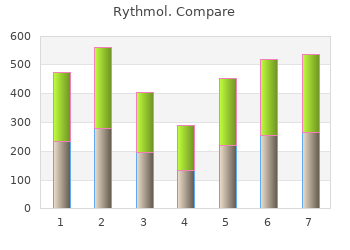
| Comparative prices of Rythmol | ||
| # | Retailer | Average price |
| 1 | Verizon Wireless | 567 |
| 2 | Alimentation Couche-Tard | 996 |
| 3 | 7-Eleven | 822 |
| 4 | Walgreen | 571 |
| 5 | Tractor Supply Co. | 298 |
| 6 | Army Air Force Exchange | 773 |
| 7 | Foot Locker | 525 |
| 8 | OSI Restaurant Partners | 275 |
| 9 | O'Reilly Automotive | 821 |
| 10 | ShopRite | 265 |
10 of 10 - Review by E. Grim
Votes: 128 votes
Total customer reviews: 128

